SBRC International Cryo-EM Seminar No.29

“Using phase diagrams with microseeding to prepare crystal samples for advanced data collection techniques”

_

Date and Time

2:00 PM – 3:00 PM Tuesday, December 9th, 2025

Location

Hybrid(CryoEM building and Zoom)


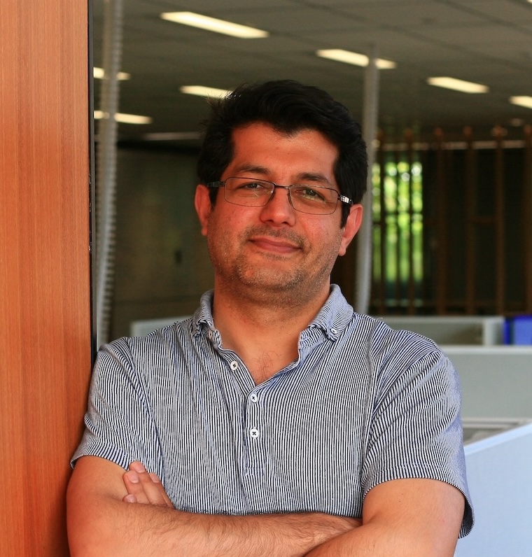
Speakers: Patrick D. Shaw Stewart

Douglas Instruments Ltd(UK)
University of Southampton(UK)


Abstract

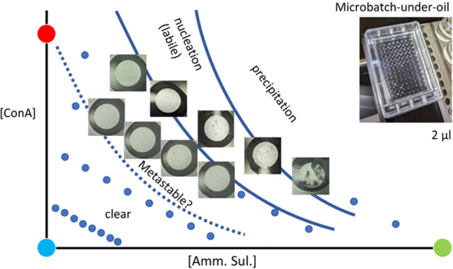
Serial data collection and microED techniques typically require “slurries” of tiny, well-ordered crystals [1]. Neutron diffraction requires very large single crystals. Creating samples for these techniques is often a complex process that requires multiple rounds of optimization. To guide them in this task, protein crystallizers often keep a notional phase diagram in mind, which has four zones: an undersaturated zone where protein always remains in solution, a metastable zone where crystals will grow when seeds are added, a crystal nucleation zone where crystals appear spontaneously, and a protein precipitation zone. However, the shape of real-life phase diagrams can vary, making the interpretation of experimental results difficult. It is therefore very helpful to determine the phase diagrams of individual target proteins experimentally. Douglas Instruments, in collaboration with the University of Southampton, has introduced a rapid and straightforward method for generating custom phase diagrams using just 15 – 60 µL of protein [Fig. 1]. The most straightforward approach utilizes the microbatch-under-oil method to prevent concentrating the sample drop (as would occur in a vapor diffusion setup). By carrying out the same procedure with and without a seedstock, the metastable zone can be identified [2]. Moreover, advanced methods often require relatively large sample volumes, and microbatch can easily be scaled up to 50 µL or larger “batches” using robotics. A new variation of the method eliminates the need for oil by using a sitting drop setup, where solutions are dispensed to the reservoirs that exactly balance the concentrations of the drops. We present case studies where phase diagrams were utilized to enhance control and crystal quality for both routine and advanced data collection.

Figure 1.  The rapid determination of a protein’s phase diagram using a microbatch-under-oil format.  Blue circles indicate the conditions that were tested.  Images of the wells are shown in conditions of interest. All points on the accessible phase diagram can be reached by mixing the three ingredients shown: protein stock (red circle), precipitant or cocktail stock (green circle) and a diluent, normally water (cyan circle).  To find the border of the metastable zone (dotted line) the experiment was repeated with the addition of a seed-stock (results not shown).

[1] Stubbs, J., Hornsey, T., Hanrahan, L.B. Esteban, R. Bolton, M.
Maly, S. Basu, J. Orlans, D. de Sanctis, J. Shim, Shaw Stewart, P. D.,
A.M. Orville, I. Tews and West, J. (2024). IUCrJ  11.

[2] D’Arcy, A., Villard, F., Marsh, M.  (2007). Acta Cryst. D63(4):550-4.


Click here to apply

【KEK-IMSS-SBRC】企業説明会

_

今年度の企業説明会は、下記日時にて開催予定とさせていただきます。

【開催日時】2025年12月12日(金)13:00〜15:00 終了予定

【会場】KEK 構造生物実験準備棟 会議室 MAP

お申込みはこちら
締め切りました


*プログラムの変更があった場合は、随時更新いたします*

【プログラム】

12月12日(金)

13:00- 13:15 構造生物学研究センターの活動について:千田俊哉


13:15- 13:30 タンパク質結晶化スクリーニングシステムについて:加藤


13:30-13:45 タンパク質結晶構造解析ビームラインについて:松垣


13:45-14:00 クライオ電子顕微鏡について:川崎


14:00-14:30 休憩(+SBRCスタッフとの交流)


14:30-14:45 100kV電顕の導入について:千田、守屋

14:45-15:00 自由討論


【 問い合わせ】 

高エネルギー加速器研究機構・物質構造科学研究所・構造生物学研究センター
引田理英
E-mail: masahide.hikita[at]kek.jp


Webページ問合せ先:増田
E-mail: cmasuda[at]post.kek.jp 

SBRC Seminar (International Cryo-EM Seminar No.28)

“Structural Studies of Viral Macromolecular Complexes by Cryo-Electron Microscopy ”

_

Date and Time

4:00 PM – 5:30 PM Friday, October 3th, 2025

Location

Onsite(CryoEM building)


Speakers: Yoko Fujita-Fujiharu

Postdoc
Max Planck Institute of Biochemistry
German,


Abstract

Understanding the architecture of viral macromolecular complexes is essential for elucidating the mechanisms of viral assembly and replication. While traditional structural biology often focuses on purified proteins in isolation, cryo-electron microscopy (cryo-EM) has the potential to analyze three-dimensional structures in more biologically relevant contexts.

In this talk, I will present some results from a study integrating multi-scale structural information of Ebola virus-like particles (VLPs). By combining in situ single-particle analysis and cryo-electron tomography, we resolved the structures of the viral matrix protein VP40 and the glycoprotein GP within intact VLPs. This multi-scale cryo-EM approach provides structural insights ranging from angstrom-level atomic detail to nanometer-scale organization, offering a more comprehensive understanding of viral assembly and function.


Click here to apply

Registration for international seminar28

Registration for

SBRC Seminar(SBRC International Cryo-EM Seminar28)

【First Name】*

【Family Name】*

【Affiliation/Company】 *

【E-mail】 *

【Participation Format】 *
This seminar will be held onsite only.

【Message】 


After registering, please confirm your registration details in the acceptance e-mail.
If you do not receive an email, your registration may not have been accepted.
Please contact us.

SBRC Seminar (International Cryo-EM Seminar No.27)

“Enzymes: From Natural Diversity to Optimized Tools for Biomedical Applications – The Case of Antimicrobial Enzymes”

_

Date and Time

10:00 AM – 11:30 AM Wednesday, September 24th, 2025

Location

Onsite(SBRC building)


Speakers: Dr. Claire STINES-CHAUMEIL

Associate Professor
Paul Pascal Research Centre
University of Bordeaux, FRANCE


Abstract

Enzymes are protein catalysts that can accelerate reaction rates by up to 1021-fold without altering the thermodynamic equilibrium. They are ubiquitous in biochemical transformations and often exhibit remarkable selectivity, a property rarely achieved by artificial catalysts. Enzymes have broad applications across various industries, including agri-food, textiles, PET degradation, drug synthesis, and medicine.
My research focuses on understanding the structure-function relationship of enzymes and elucidating their mechanisms, particularly those relevant to biomedical and biotechnological applications. I then optimize natural catalysts through genetic and protein engineering to enhance their suitability for specific uses, such as glucose biosensors, catheters, immunosensors, and disinfection.
In my talk, I will focus on microbicidal enzymes naturally involved in human innate immunity. I will present the strategies used to identify key enzymes and characterize their biochemical and physicochemical properties. These enzymes have been optimized to exhibit ideal antimicrobial properties for biomedical applications, such as combating nosocomial infections.


Click here to apply

SBRC International Cryo-EM Seminar No.26

Time-resolved cryo-EM reveals mechanism of allosteric activation in isocitrate lyase 2

_

Date and Time

4:00 PM – 5:30 PM Friday, September 26th, 2025

Location

Hybrid (Zoom and KEK CryoEM building)


Speakers : Ghader Bashiri


Associate Professor
Laboratory of Microbial Biochemistry and Biotechnology
School of Biological Sciences, The University of Auckland

Abstract

Allostery is a fundamental regulatory mechanism underpinning many biochemical and physiological processes within cells. Allostery occurs when a binding event at one site on a protein affects binding at a remote functional site, thereby enabling precise control of enzymatic function. Isocitrate lyase 2 (ICL2) serves as a regulatory hub in the central metabolism of Mycobacterium tuberculosis, the bacterium that causes tuberculosis, where is modulates carbon flux in the central carbon metabolism.

Our crystal structures of Mtb-ICL2 demonstrate that binding of acetyl-CoA or propionyl-CoA at a site distant to the active site induces extensive conformational changes, leading to a ~100-fold increase in its enzymatic activity. Using time-resolved cryo-EM, we captured the trajectory of these structural transitions, from a time point as early as 150 ms through to the fully active state. Our data reveal the dynamic population shifts between protein conformations over time. Our findings provide unprecedented insights into the mechanisms underlying allosteric activation of Mtb-ICL2.


Click here to apply

SBRC Seminar (International Cryo-EM Seminar No.25)

“医学はアート?〜サイエンスに基づく臨床の思考プロセスに触れる〜”

_

Date and Time

2025年8月29日(金) 10:00

Location

オンサイト(構造生物棟会議室)


Speakers:

戒能 賢太

筑波大学 医学医療系 内分泌代謝・糖尿病内科


Abstract

構造生物学を含むあらゆる生物学研究の中でも、特に哺乳類を対象とした研究は、臨床応用を目的とすることが多いかと思います。研究成果に基づく診断ツールとしての検査法の開発や、新たな治療法の創出は比較的イメージしやすく、実際に日々新しい検査法や治療法が臨床の場で応用されており、医学は常に進歩を遂げていると言えます。しかし「検査」や「治療」といった選択肢は、患者さんが病院を訪れてから何らかの転帰に至るまでの過程全体の中ではあくまで一部分に過ぎず、実際、臨床医の思考の多くは適切な検査や治療を選択するまでのプロセスに費やされています。医学教育の礎を築いたウィリアム・オスラーは、そのプロセスを指して「医学とは、科学に基づく技術(アート)である」と表現しましたが、今回のセミナーでは、医学が対象とする領域の中でもそうした「アート的」側面に焦点を当て、その一端に触れていただければと思います。


Click here to apply

SBRC Seminar

Structural Studies of Biomolecular and Supramolecular Complexes

_

Date and Time

4:00 PM – Friday, August 22, 2025

Location

KEK CryoEM building


Speakers: Ilkin Yapici

Koc University

Abstract

High-resolution structural studies continue to drive progress in both antibiotic development and neurodegenerative disease research. Building on our previous work using serial femtosecond X-ray crystallography (SFX) at XFELs to resolve 30S and 50S ribosomal subunits at ambient temperature, we are now extending our structural investigations to intact 70S ribosomes using single-particle cryo-electron microscopy. These cryo-EM studies aim to elucidate how next-generation antibiotics interact with the bacterial translation machinery, offering new insights into drug binding, resistance mechanisms, and ribosome functional dynamics under near-native conditions.

In parallel, we are investigating the structural features of ALS-linked superoxide dismutase 1 (SOD1) aggregates, with a focus on the H71Y mutant identified in Turkish patients. Cryo-EM and cryo-electron tomography (cryo-ET) reveal that this variant forms long, unbranched amyloid-like fibers with a characteristic hollow-core morphology and accelerated aggregation kinetics. Guided by these structural insights, we are screening a panel of hybrid small molecules incorporating radical-scavenging, metal-chelating, and ferroptosis-inhibiting elements. Preliminary data suggest these compounds may interfere with fiber growth, destabilize preformed filaments, or inhibit pathological seeding.

Together, these efforts illustrate how complementary high-resolution techniques, XFEL-based SFX and cryo-EM, can be leveraged to investigate both essential biomolecular complexes and disease-relevant protein assemblies. Our findings contribute to structure-guided strategies for the development of new antibiotics and therapeutic approaches for ALS.


Click here to apply

SBRC International Cryo-EM Seminar No.24

“DeepMASC: Automated Mask Creation and Class Selection in Single Particle Analysis Empowered by Deep Protein Probability Measures of Cryo-EM Maps”

_

Date and Time

4:00 PM – 5:30 PM Monday, June 16th, 2025

Location

Hybrid (Zoom and KEK SBRC building)


Speakers:

Mr. Han Zhu, M.S.

Department of Computer Science, Purdue University, US.


Abstract

We have developed DeepMASC (Deep Masking and Auto-Selection for Classifications), a novel approach that eliminates human intervention from two critical steps in the cryo-EM single particle analysis (SPA) computational workflow: 3D class selection and 3D reference mask generation. Our method leverages deep learning-based protein probability measures to automatically identify optimal structural classes and generate appropriate masks for further refinement. DeepMASC dramatically accelerates the SPA workflow by eliminating time-consuming manual interventions that often create significant delays. Traditional workflows require expert decisions at critical junctures, creating bottlenecks that can extend processing time from hours to days. By automating these decision points, DeepMASC enables continuous, uninterrupted processing without sacrificing quality.
To address broader computational challenges in cryo-EM analysis, we also have developed the Kihara Lab EMSuite Server, a free and comprehensive web platform offering 13 algorithms for cryo-EM analysis. The server spans the full spectrum of needs, from secondary structure detection at medium resolution to atomic modeling at high resolution for proteins, nucleic acids, and their complexes. This unified interface makes sophisticated cryo-EM analysis accessible to structural biologists regardless of computational expertise, democratizing access to cutting-edge methods without requiring specialized hardware or software installation.

Profile

2018-2022              B.S., Department of Computer Science, Purdue University, US.

2022-2023              M.S., Department of Computer Science, Purdue University, US.

2023-Present          Ph.D. program, Department of Computer Science, Purdue University, US

Research Focus

Computational Structural Biology, ML-Based Cryo-EM Modeling and Analysis


Click here to apply